
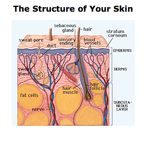
The structure of your skin. Your skin’s anatomy, can be divided into two main layers, the epidermis and dermis. The uppermost layer of the skin structure is the epidermis, which itself contains several layers — the basal cell layer, the spinous cell layer, the granular cell layer, and the stratum corneum. The deepest layer of the epidermis is the basal cell layer. Here cells divide to produce new skin cells. These cells move towards the skin surface, pushed upward by the dividing cells below them.
Blood vessels in the dermis — which is below the basal cell layer — supply nutrients to support this active growth of new skin cells.
The stratum corneum is the top layer of the epidermis — it is the layer of the skin that we see from the outside. These cells are dead, contain a lot of keratin and are arranged in overlapping layers that impart a tough and waterproof character to the skin’s surface.
Dead skin cells are continually shed from the skin’s surface. This is balanced by the dividing cells in the basal cell layer to produce a state of constant renewal. Also in the basal cell layer are cells called melanocytes that produce melanin. Melanin is a pigment that is absorbed into the dividing skin cells to help protect them against damage from sunlight (ultraviolet light). The amount of melanin in your skin is determined by your genes and by how much exposure to sunlight you have. The more melanin pigment present, the darker the colour of your skin.
The epidermis also contains dendritic cells, which are part of the immune system and help protect the body from foreign substances.
The dermis contains a variable amount of fat, and also collagen and elastin fibres which provide strength and flexibility to the skin.
When the skin is exposed to sunlight, modified cholesterol in the dermis produces vitamin D, which helps the body to absorb calcium for healthy bones.
Blood vessels supply nutrients to the dividing cells in the basal layer and remove any waste products. They also help maintain body temperature by dilating and carrying more blood when the body needs to lose heat from its surface; they narrow and carry less blood when the body needs to limit the amount of heat lost at its surface.
Nerves in the dermis detect heat, cold, pain, pressure and touch and relay this information to the brain. In this way the body senses changes in the environment that may potentially harm the body.
A sebaceous gland opens into each hair follicle and produces sebum, a lubricant for the hair and skin that helps repel water, damaging chemicals and microorganisms.
Sweat glands occur on all skin areas — each person has more than 2 million. When the body needs to lose heat these glands produce sweat. Sweat moves to the skin’s surface via the sweat duct, and evaporation of this water from the skin has a cooling effect on the body.
The innermost layer of the skin structure is the layer of subcutaneous fat, and its thickness varies in different regions of the body. The fat stored in this layer represents an energy source for the body and helps to insulate the body against changes in the outside temperature.
Despite this complex structure, modern dermatological medicine tends to see skin lesions as purely local affections, or at least usually treat them as such. Thus suppressive treatments like cortisone are applied directly to the eruption in order to make it ‘go away’.